axon terminal svg
+465 axon terminal svg cut files vector Files from our Design Marketplace.
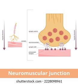
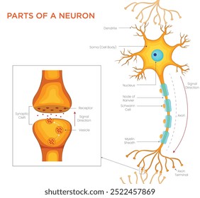
Neuromuscular junction. A synaptic connection between the terminal end of a motor nerve and a muscle. Presynaptic (nerve terminal), postsynaptic part, synaptic cleft.
Synaptic Cleft Axon Terminal science vector illustration graphic template
An axon terminal is transmitting a signal to neuron.Vector illustration.
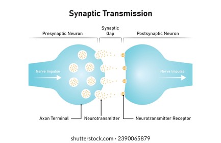
Synaptic Transmission Scientific Design. Vector Illustration.
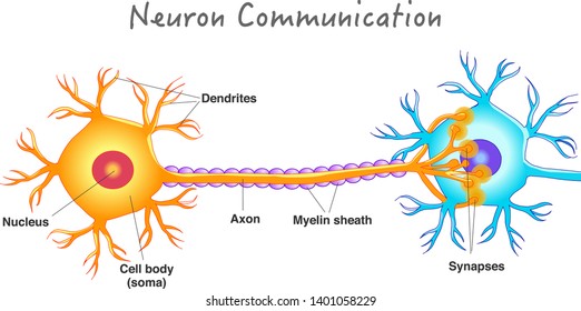
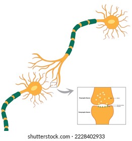
Neuron communication. Transmission of the nerve signal between two neurons. Neuron connect. Nervous system. Simple annotated. White background. 2d vector drawing.
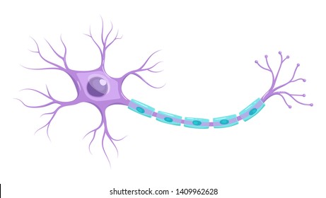
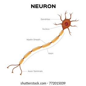
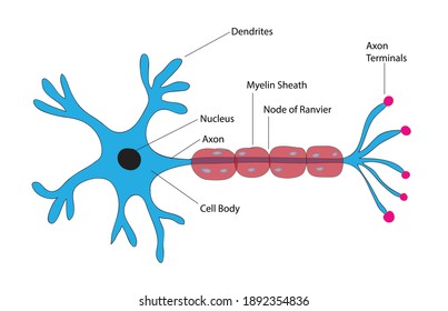
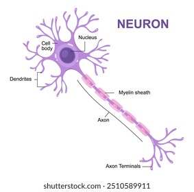
Illustration of neuron anatomy. Vector infographic (nerve cell axon and myelin sheath)
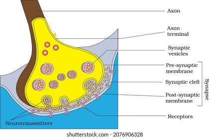
Diagram showing axon terminal and synapse
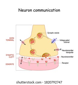
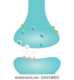
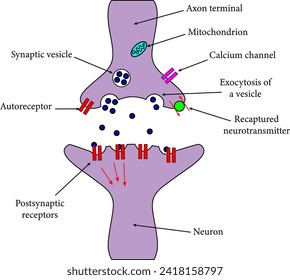
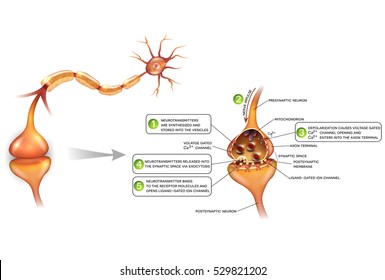
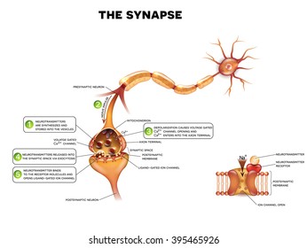
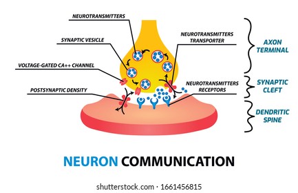
Chemical synapse structure. Neuron communication. Close-up of axon terminal with Synaptic vesicles, Voltage-gated channel, and Neurotransmitter transporters. neurotransmitter binds to receptor
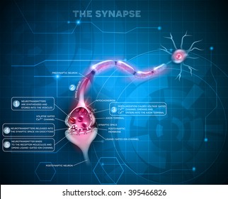
Synapse detailed anatomy on an abstract technology background. Neuron passes signal to another
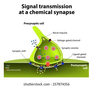
Signal transmission at a chemical synapse. one neuron releases neurotransmitter molecules into a synaptic cleft that is adjacent to another neuron.
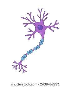
Structure of a motor neuron. Anatomy of the neuron of the brain
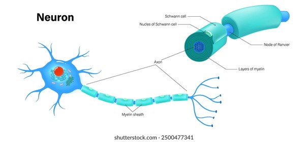
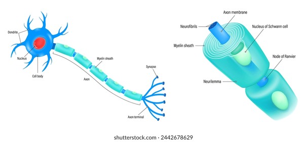
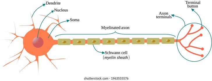
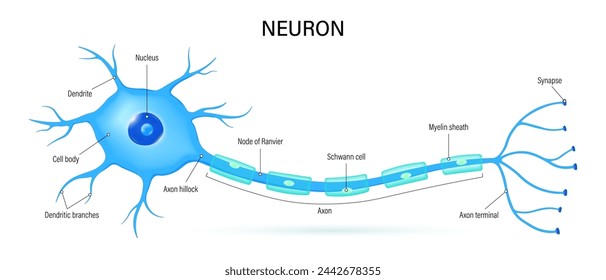
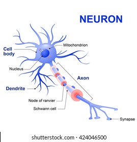
Neuron and components of the Myelin sheath anatomy vector. Cell body, dendrite, Axon, Synapse, myelin sheath, node Ranvier and Schwann cell.
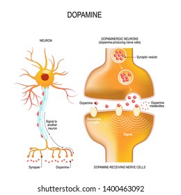
Dopamine. closeup presynaptic axon terminal, synaptic cleft, and dopamine-receiving nerve and dopamine-producing cells. Labeled diagram. Vector illustration for educational, biological, medical use

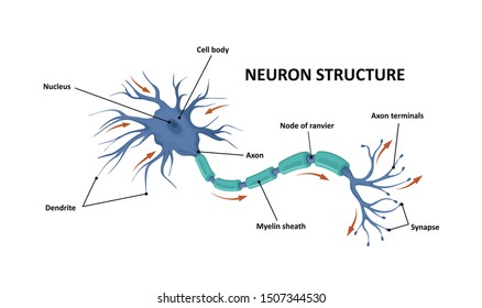
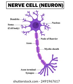
A colorful illustrated neuroscience diagram displaying a neuron cell with labeled dendrites, soma, nucleus, axon hillock, myelin sheath, nodes of Ranvier, and axon terminals highlighting action potent
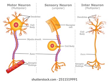
Neuron types, motor, sensory, inter. Different nerve cells, multipolar, unipolar anatomy. Parts, axon, body, dendrites connect, myelin sheath. junction transmission structures. Vector illustration
Neurons and closeup of synapse detailed anatomy, beautiful colorful illustration. Neuron passes signal to another neuron.
how neurotransmitter works? The process the brain neurons communicate each other anatomy vector illustration. Action potentials and synapses. multiple sclerosis formation. Motor neuron communication.
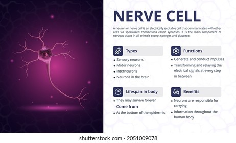
Structure, Function and Types of Nerve Cell Vector Image Design
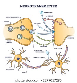
Neurotransmitter process detailed anatomical explanation outline diagram. Labeled educational scheme with vesicle, axon terminal, enzyme production and receptors vector illustration. Synapse impulse.
Vector Illustration of neuron anatomy (nerve cell axon and myelin sheath
Neuron and components of the Myelin sheath vector. Anatomy of a typical human neuron. Cell body, dendrite, Axon, Synapse, myelin sheath, node Ranvier and Schwann cell.
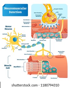
Neuromuscular junction vector illustration scheme. Labeled medical infographic. Motor neuron and muscle cell structure closeup. Diagram with myofibril and muscle fibers.
Vector Illustration of neuron anatomy. Doodle style (nerve cell axon and myelin sheath)

Diagram of Neuron Anatomy illustration
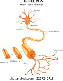
Colorful Neuron anatomy and myelin sheath formation on a white background
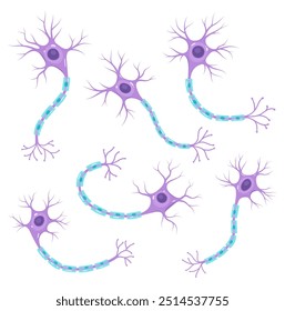
Vector set of neurons. Scientific illustration
Structure of a motor neuron.Anatomy of nerve cell.Nervous system.Axon, dendrites, myelin sheath and soma.Diagram for for biology.Education Chart.Schwann cell.Infograhic.Cartoon vector illustration.
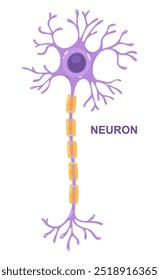
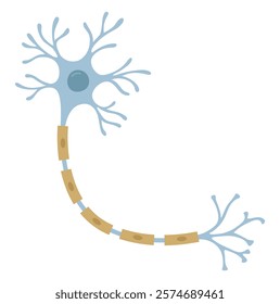
Neuron that is the main part of the nervous system.
Neuron anatomy vector . infographic
Vector infographic of neuron anatomy. Axon, myelin sheat, dendrites, cell body, nucleus.
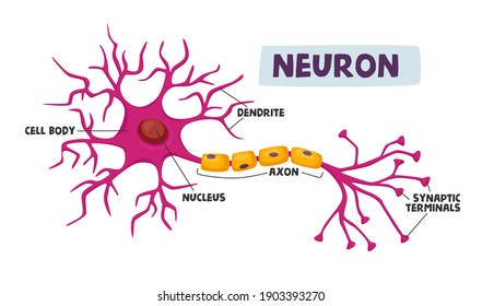
Human Neurons Scheme Infographics Dendrite, Cell Body, Axon and Nucleus with Synaptic Terminals Scientific Medical Infographic, Learning Aid Isolated on White Background. Cartoon Vector Illustration
Neuron Synapse illustration. Conection between pre and pos synaptic neuron illustration
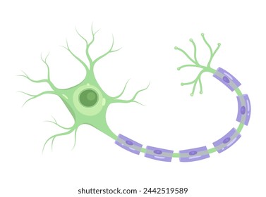
Multipolar neuron with myelinated axon
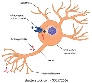
Development of an action potential in a nerve cell through the action of a voltage-gated sodium channel in the cell body.
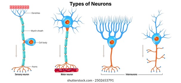
Types of neurons vector. Sensory neuron, Motor neuron and Interneurons structure. Neuron types. Nerve cell anatomy.
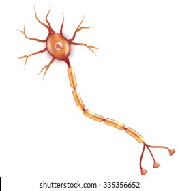
Neuron vector illustration Isolated on a white background.

Nerve impulse traveling through axon terminal and synapse
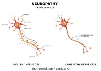
Neuropathy that is the damage of nerves, this can be caused by Diabetes.
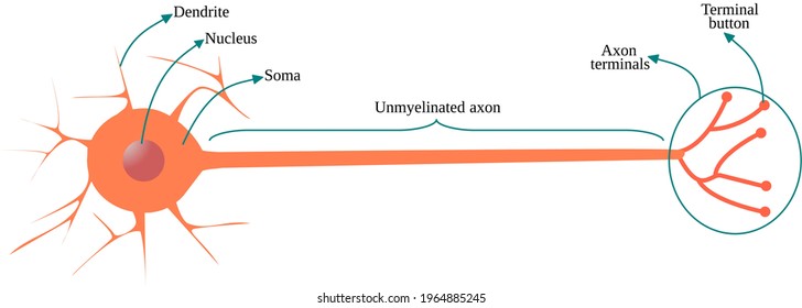
Multipolar neuron with unmyelinated axon
Biological anatomy of typical neuron cell, detailed neurone cells, detailed neuron cell, basic structure of typical neuron cells, Human nerve cell, cell body (soma), dendrites, and a single axon

Axon. Neurites are nerve cells that carry nerve impulses from the soma to innervated organs and other nerve cells.

Structure of a motor neuron, vector illustration
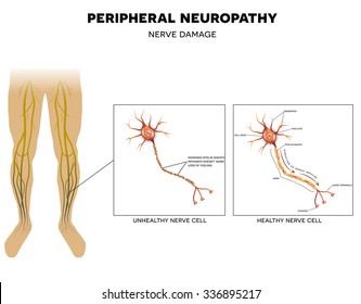
Neuropathy that is the damage of peripheral nerves that causes pain and loss of sensation in the extremities.
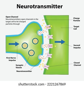
Neurotransmitters are released from synaptic vesicles of the presynaptic neuron and bind to receptors on the postsynaptic neuron, triggering an impulse through the 2nd neuron.
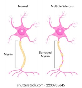
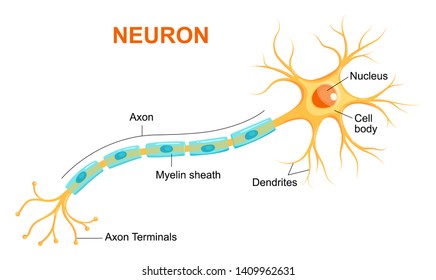
Neuron concept vector. Dendrite, axon, soma of neuron. Multiple sclerosis, nerve anatomy illustration. Myelin and nucleus of brain cell.
Synapse detailed anatomy, beautiful colorful illustration. Neuron passes signal. At the right side closer look at Ligand gated ion channel.
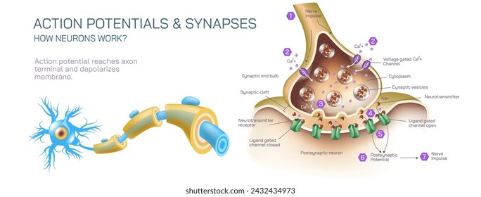
how neurotransmitter works? The process the brain neurons communicate each other anatomy vector illustration. Action potentials and synapses. multiple sclerosis formation. Motor neuron communication.
Neuron anatomy vector. Nerve cell diagram. Axon, dendrites and synapse.

Neural connection diagram. Structure of neuron with axon, dendrites and soma. Transmission of nerve impulse or electrical signal across synapse.
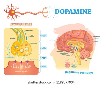
Dopamine vector illustration. Labeled diagram with its action and pathways. Scheme with closeup presynaptic axon, terminal, synaptic cleft, dendrite and receiving cells.
A neuron consists of a cell body (soma), dendrites, axon, myelin sheath, axon terminals, and synapse, enabling communication within the nervous system.
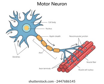
Human anatomy of a motor neuron, including its parts like the axon and dendrites structure diagram hand drawn schematic vector illustration. Medical science educational illustration
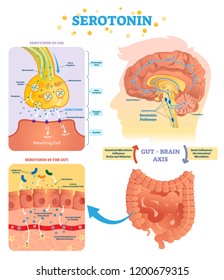
Serotonin vector illustration. Labeled diagram with gut brain axis and CNS. Intestinal microbiota influence brain behavior and intestinal cycle. Educational infographic.
Vector Illustration of neuron anatomy. Nerve cell axon and myelin sheath
Neuron line icon. Nerve cell showing axon, dendrites, and cell body. Brain cells, synapses, nervous system, and communication between neurons. Neuroscience, psychology, medicine. Vector illustration
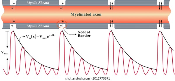
Saltatory conduction: way an electrical impulse skips from node to node down the full length of an axon
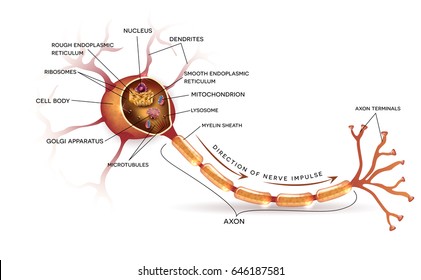
Anatomy of a typical human neuron

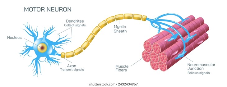
Educational vector of labeled healthy motor neuron, showing axon, myelin sheath, and muscle connection.
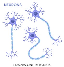
Vector set of neurons. Scientific illusteration
Neuron, nerve cell that is the main part of the nervous system. Cross section detailed anatomy, nucleus and other organelles of the cell.
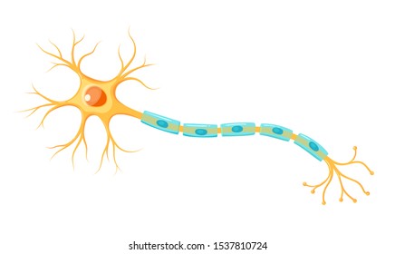
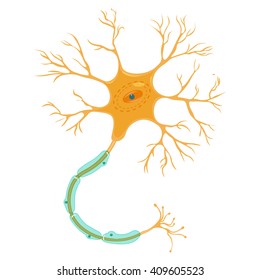
Vector yellow neuron isolated on white background. Educational illustration
Vector Illustration of neuron anatomy. Scientific infographic (nerve cell axon and myelin sheath)

Human nerve cell red translucent low poly triangles on dark blue background. Futuristic glowing organ hologram and copy space for text. Medical and science concept. Banner design vector.
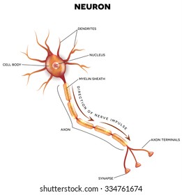
Neuron that is the main part of the nervous system with description
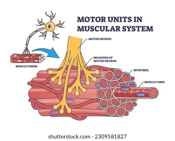
Motor units in muscular system with fibers neuron anatomy outline diagram. Labeled educational medical scheme with myofibril and muscle fiber closeup vector illustration. Nerve functional contraction
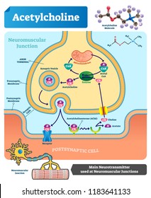
Acetylcholine vector illustration. Labeled scheme with structure of neurotransmitter, neuromuscular junction, synaptic vesicle, axon and cleft. Anatomical closeup diagram
Vector infographic of neuron anatomy. Axon, myelin sheat, dendrites, cell body, nucleus.
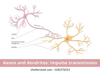
Axons and dendrites: impulse transmission. Nerve cells and messages between them. Visualization of human neurological system communication. Electrochemical stimulation received from neural cells.

Anatomy of a typical human neuron (axon, synapse, dendrite, mitochondrion, myelin sheath, node Ranvier and Schwann cell). Vector diagram
Neuron communication concept. Axon terminal, synaptic cleft, dendritic spine. Vector illustration.